The Most Posterior Aspect of a Typical Vertebra Is the
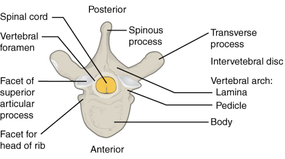
Vertebral arch - the structure located posterior to the body. The arch along with the posterior aspect of the body forms the vertebral spinal canal which contains the spinal cord.
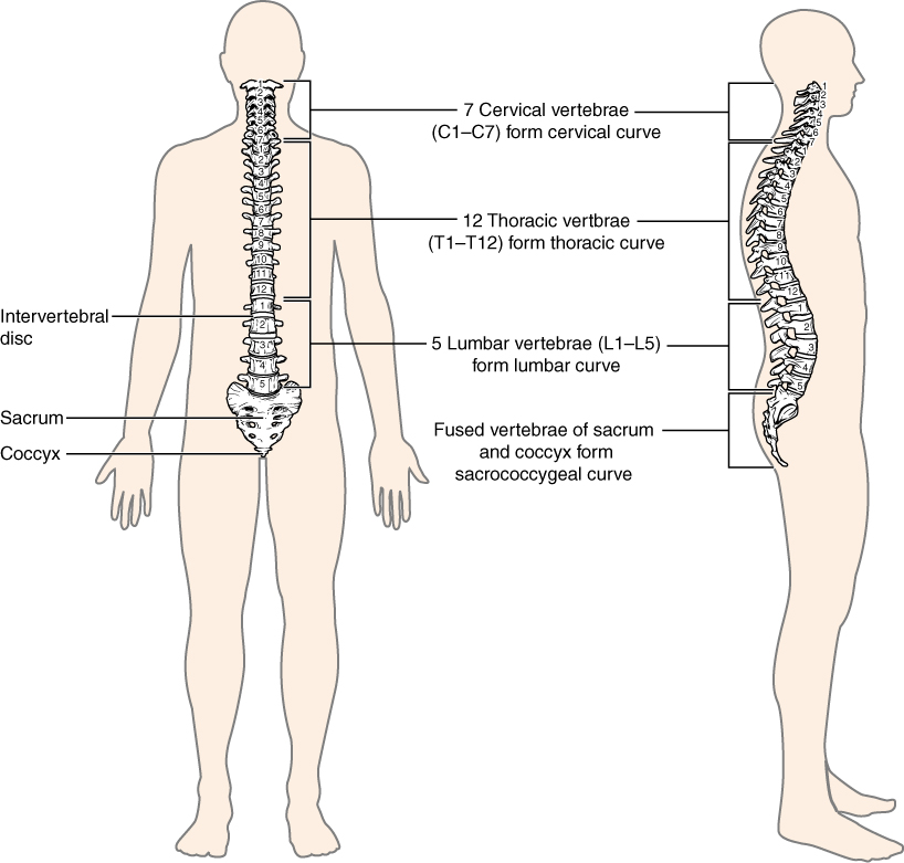
The Vertebral Column Anatomy And Physiology
Hemangiomas of the vertebral bodies are common benign vascular tumors.

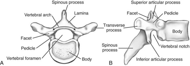
. The arch consists of bilateral pedicles pieces of bone that connect the arch to the body and bilateral lamina bone segments form most of the arch connecting the transverse and spinous processes. The most posterior aspect of a typical vertebra is the. September 19 2012 by gvs14 in Vertebrae.
Each pedicle forms one of the lateral sides of the vertebral arch. Each pedicle forms one of the lateral sides of the vertebral arch. The arch consists of bilateral pedicles cylindrical segments of bone that connect the arch to the body and bilateral lamina bone segments form most of the arch connecting the transverse and spinous processes.
1 Most of the lesions occur in the thoracolumbar spine. The posterior arch of the atlas forms the posterior three-fifths of the circumference of the atlantal ring and is homologous with the pedicles and laminae of typical vertebrae. The inferior aspect of the sacrum articulates with the coccyx via the sacral and coccygeal cornua.
The bony structures connected directly to the vertebral body are the. The arch along with the posterior aspect of the body forms the vertebral spinal canal which contains the spinal cord. The only atypical vertebra of the lumbar region is L5.
The arch consists of bilateral pedicles pieces of bone that connect the arch to the body and bilateral lamina bone segments form most of the arch connecting the transverse and spinous processes. It serves as the attachment point for the supraspinous ligament. L5 also has the most inferiorly located discovertebral unit in the human vertebral column.
The median sacral crest forms from the fusion of the first three sacral spinous processes. The pedicles are anchored to the posterior side of the vertebral body. It serves as the attachment point for the supraspinous ligament.
The inferior aspect of the sacrum articulates with the coccyx via the sacral and coccygeal cornua. These facilitate the passage of spinal nerves from the spinal cord. The posterior aspect of the sacrum exhibits numerous bony landmarks.
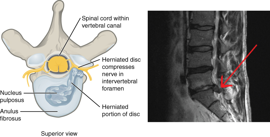
Coursing through the intervertebral foramen with the vertebral arteries the. Each vertebra articulates with the vertebrae above and below it via an intervertebral disc. The majority of the hemangiomas seen with imaging studies are asymptomatic and incidental findings.
The neural arch is comprised of the bone posterior to the vertebral body which has several individual components that are fused to form a ring the vertebral foramen that encloses the spinal canal. Typical Vertebra Features. The pedicles contain vertebral notches superior inferior which form intervertebral foramina.
On its sides it has semi-ovular indentations for ribs called costal facets not shown. The arch along with the posterior aspect of the body forms the vertebral spinal canal which contains the spinal cord. The anterior aspect of the vertebral body is higher than the posterior aspect contributing to the slightly wedge-shaped appearance it has.
The posterior aspect of the sacrum exhibits numerous bony landmarks. The median sacral crest forms from the fusion of the first three sacral spinous processes. The arch consists of bilateral pedicles cylindrical processes of bone that connect the arch to the body and bilateral lamina flat bone segments form most of the arch connecting the transverse and spinous processes.
Abnormal increased convexityof the spine. The arch along with the posterior aspect of the body forms the vertebral spinal canal which contains the spinal cord. The pedicles are anchored to the posterior side of the vertebral body.
Vertebral body hemangiomas are the most common tumor of the spinal axis and occur in approximately 10-20 of adults. The superior surface of the posterior arch bears a groove which is simply known as the groove for the vertebral artery. The vertebral arch forms the posterior portion of each vertebra.
L5 has the largest vertebral body and transverse processes. The arch consists of bilateral pedicles pieces of bone that connect the arch to the body and bilateral lamina bone segments form most of the arch connecting the transverse and spinous processes. The vertebral arch forms the posterior portion of each vertebra.
The arch along with the posterior aspect of the body forms the vertebral spinal canal which contains the spinal cord. Meanwhile the posterior aspect of the cervical spine receives innervation by the medial branch of the posterior primary rami. The large cylindrical portion anterior to other features.
It consists of four parts the right and left pedicles and the right and left laminae. Despite displaying most typical vertebral hallmarks a significant amount of anatomical variation exists within the cervical spine. The arch along with the posterior aspect of the body forms the vertebral spinal canal which contains the spinal cord.
It consists of four parts the right and left pedicles and the right and left laminae. The features that encircle the vertebral foramen with the body. The joints between articular processes of vertebra are termed.
The arch consists of bilateral pedicles cylindrical processes of bone that connect the arch to the body and bilateral lamina flat bone segments form most of the arch connecting the transverse and spinous processes. It consists of two pedicles and two laminae.
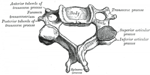
Cervical Vertebrae Physiopedia
Spine Radiology Key

Crossfit Basic Structure Of The Vertebrae
The Vertebral Column Unit 1 Flashcards Quizlet

Vertebrae An Overview Sciencedirect Topics
![]()
Vertebral Column Anatomy Vertebrae Joints Ligaments Kenhub

The Vertebral Column Anatomy And Physiology

Clinically Applied Anatomy Of The Vertebral Column Surgery Oxford International Edition

Typical Vertebral Structure Medical Coding Human Anatomy And Physiology Science Revision

Clinically Applied Anatomy Of The Vertebral Column Surgery Oxford International Edition

The Vertebral Column Anatomy And Physiology
The Vertebral Column Anatomy And Physiology
The Vertebral Column Bones Of The Spine Geeky Medics

Vertebral Column Flashcards Quizlet

Features Of Typical Vertebra Fall 2020 Msu Mediaspace

Lumbar Vertebrae An Overview Sciencedirect Topics

Structure Of A Typical Vertebrae Diagram Quizlet

Anatomy Of The Vertebral Column Surgery Oxford International Edition

Back Basicmedical Key


Comments
Post a Comment